Skeleton X Ray Biology Diagrams Normal chest x ray. Radiological anatomy is where your human anatomy knowledge meets clinical practice. It gathers several non-invasive methods for visualizing the inner body structures. The most frequently used imaging modalities are radiography (X-ray), computed tomography (CT) and magnetic resonance imaging (MRI).X-ray and CT require the use of ionizing radiation while MRI uses a magnetic
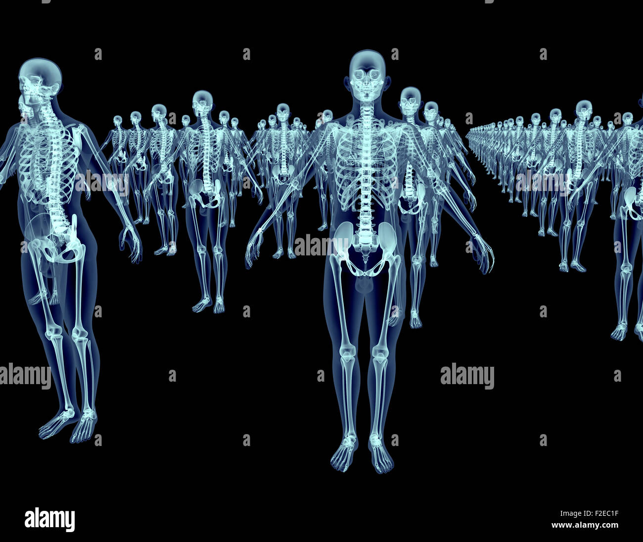
The skeleton is the complete set of bones in the human body. Traditionally there are said to be 206 bones in total which can be divided into: axial skeleton. appendicular skeleton. NB: the total of 206 bones treats the patellae as proper bones and not sesamoids, and ignores all the other sesamoids! (as well as multiple anatomic variants) Anatomy and Physiology. The skeletal system is composed of 206 separate bones and is responsible for body support, protection, movement, and blood cell production. Radiographic evaluation will demonstrate multiple fractures in various stages of healing and a general decrease in bone mass. The bone cortex is thin and porous, and the
Anatomy, the Anatomy of Imaging Biology Diagrams
This article lists a series of labeled imaging anatomy cases by body region and modality. Brain CT head: non-contrast axial CT head: non-contrast coronal CT head: non-contrast sagittal CT head: non-contrast axial with clinical questions CT X-ray interpretation (ABCS approach) The ABCS approach of X-ray interpretation involves assessing the following:. Alignment and joint space; Bone texture; Cortices; Soft tissues; General points. Don't forget to review all views, compare both sides and re‐examine any previous imaging.. If you spot one abnormality, do not lose focus until you have reviewed all areas of the image, otherwise e-Anatomy is a high-quality anatomy and imaging content atlas.It is the most complete reference of human anatomy available on the Web, iPad, iPhone and Android devices. Explore detailed anatomical views and multiple modalities (over 8,900 anatomic structures and more than 870,000 translated medical labels) with images in CT, MRI, radiographs, anatomical diagrams and nuclear images.
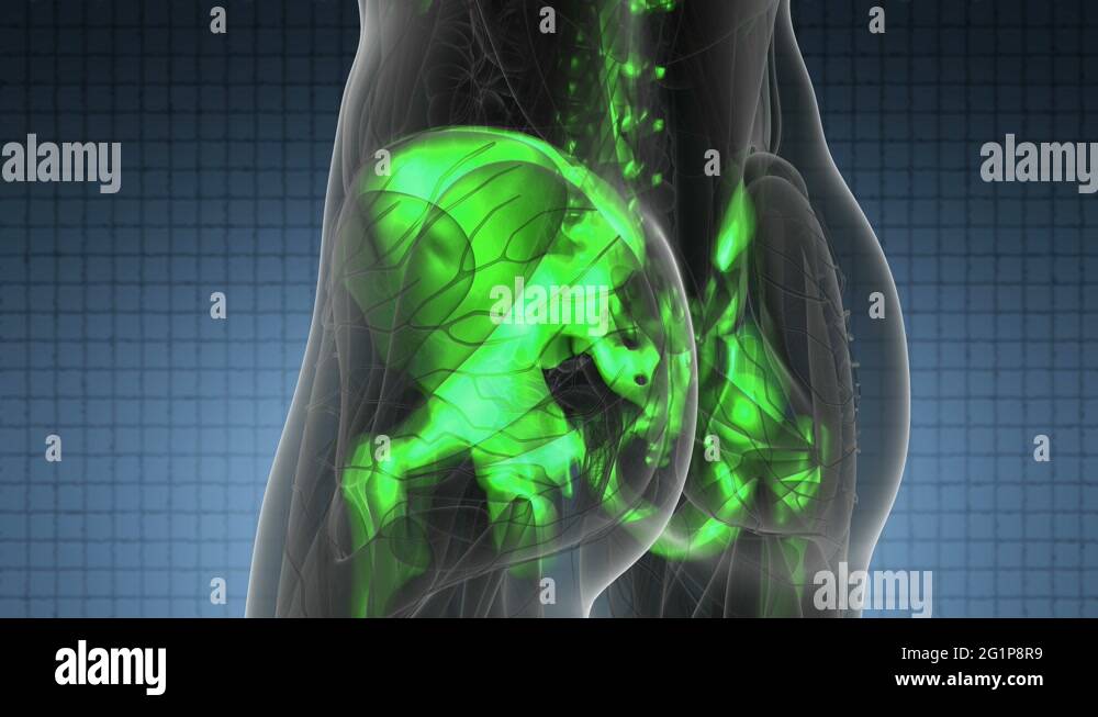
A typical skeletal survey using conventional x-ray includes bilateral anteroposterior (AP) and posteroanterior (PA) projections of hands, forearms, humerus, feet, leg, femur, pelvis, spine and skull. A joint survey includes bilateral AP and PA views of wrist, elbow, shoulder, ankle, knee, hip and sacroiliac joints. Bone anatomy example - Knee. Hover on/off image to show/hide findings. Tap on/off image to show/hide findings. Click image to align with top of page. Bone anatomy example - Knee. Long bones comprise diaphysis, metaphysis and epiphysis; The growth plate separates the metaphysis from the epiphysis until fusion in adult life